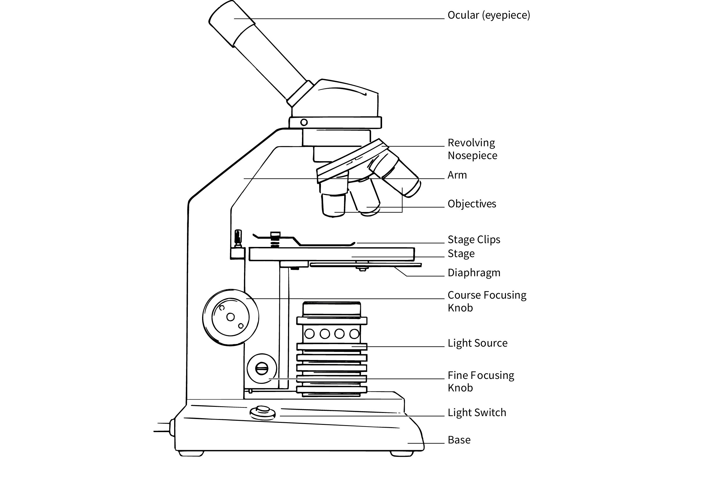
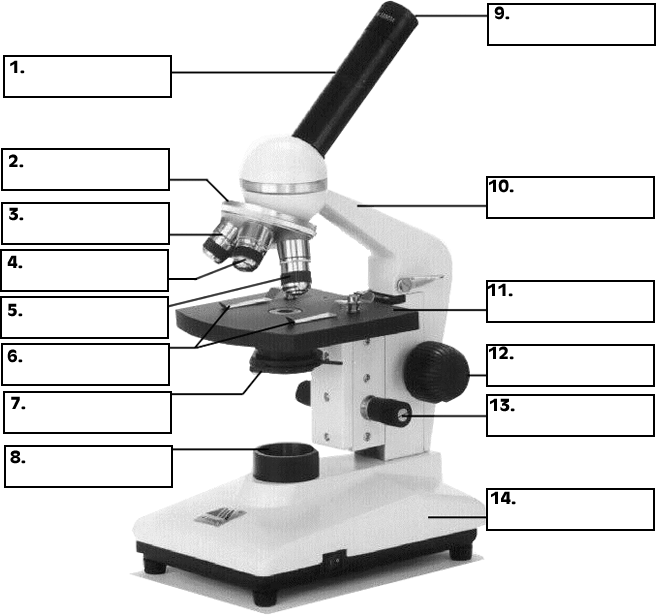
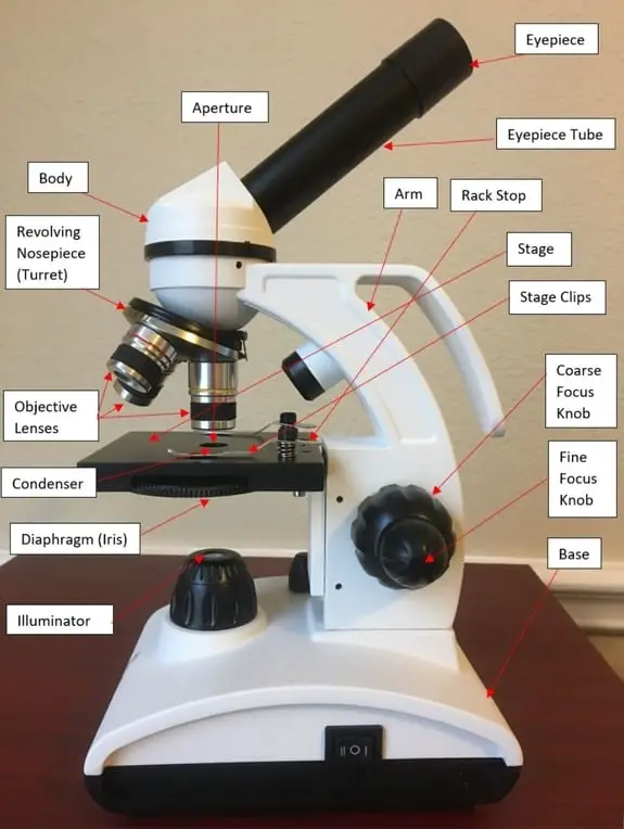
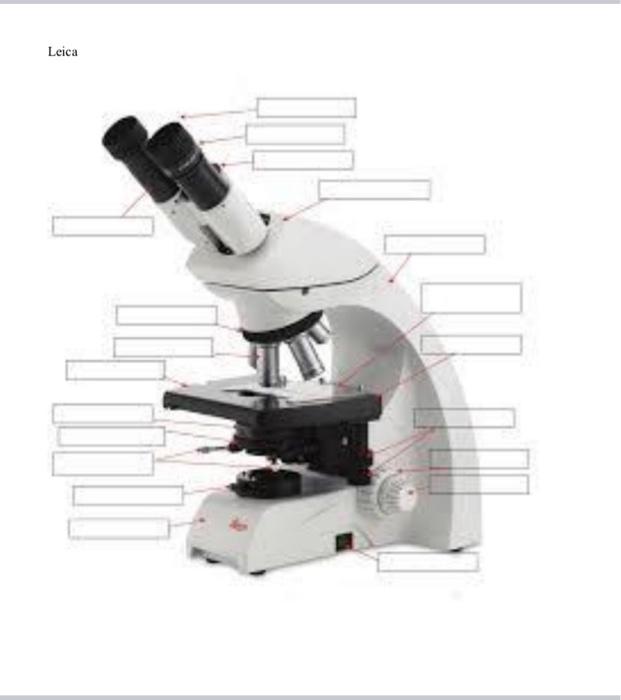
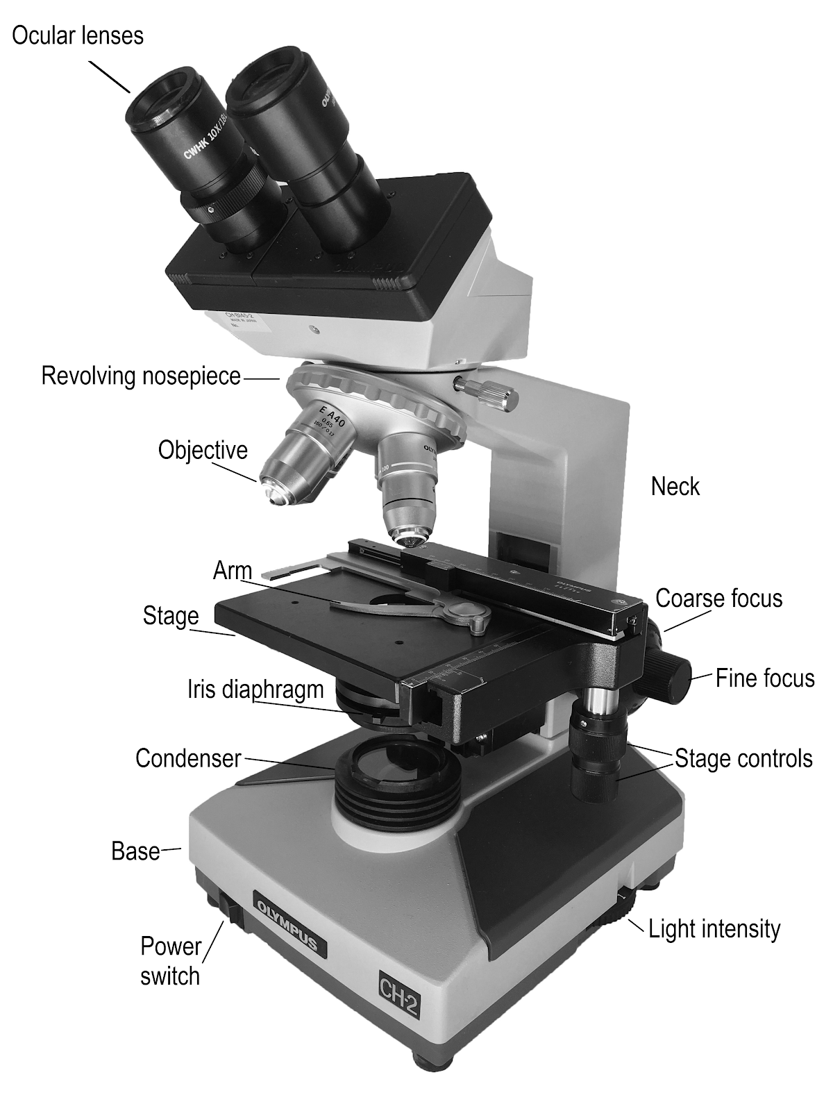
41 picture of compound microscope with labels
Chloroplast - Wikipedia A chloroplast / ˈ k l ɔːr ə ˌ p l æ s t,-p l ɑː s t / is a type of membrane-bound organelle known as a plastid that conducts photosynthesis mostly in plant and algal cells.The photosynthetic pigment chlorophyll captures the energy from sunlight, converts it, and stores it in the energy-storage molecules ATP and NADPH while freeing oxygen from water in the cells. Unbanked American households hit record low numbers in 2021 Oct 25, 2022 · The number of American households that were unbanked last year dropped to its lowest level since 2009, a dip due in part to people opening accounts to receive financial assistance during the ...
Microsoft takes the gloves off as it battles Sony for its ... Oct 12, 2022 · Microsoft pleaded for its deal on the day of the Phase 2 decision last month, but now the gloves are well and truly off. Microsoft describes the CMA’s concerns as “misplaced” and says that ...
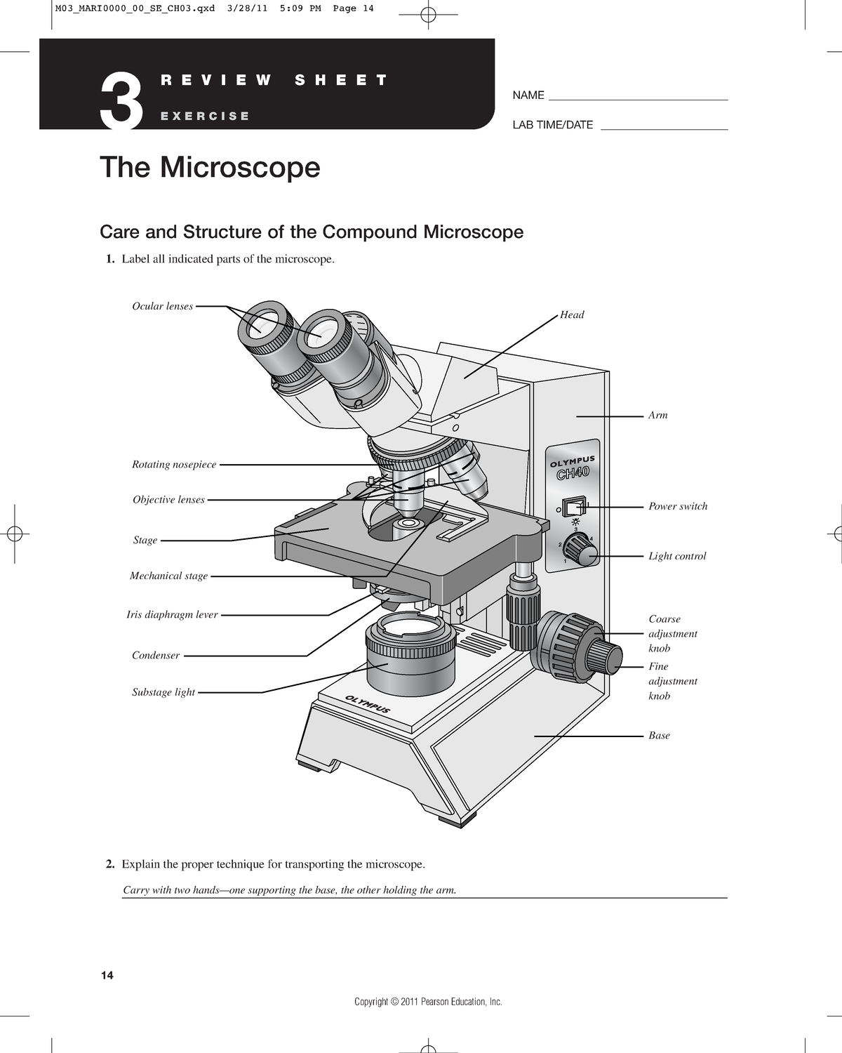
Picture of compound microscope with labels
National Geographic Dual LED Student Microscope Aug 07, 2017 · Vanstarry Beginners Microscope Kit 40X-1000X for Kids & Students, Dual LED Lights and Cordless Capability, Illumination Lab Compound Monocular Microscopes with Optical Glass Lenses & 12 Slides 4.4 out of 5 stars 116 Live Cell Imaging | Solutions | Leica Microsystems Jun 01, 2022 · To perform successful live-cell imaging experiments, using the right platform is critical. When choosing an optical microscope for live‐cell imaging, the following 3 variables should be considered: detector sensitivity (signal‐to‐noise ratio), specimen viability, and image-acquisition speed. Scanning electron microscope - Wikipedia History. An account of the early history of scanning electron microscopy has been presented by McMullan. Although Max Knoll produced a photo with a 50 mm object-field-width showing channeling contrast by the use of an electron beam scanner, it was Manfred von Ardenne who in 1937 invented a microscope with high resolution by scanning a very small raster with a demagnified and finely focused ...
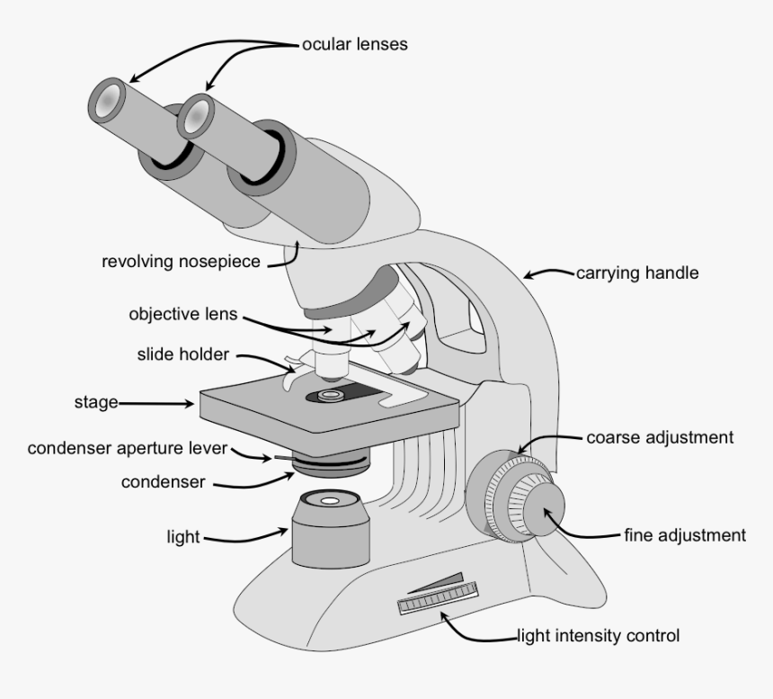
Picture of compound microscope with labels. Micro Module 1 Flashcards | Quizlet Study with Quizlet and memorize flashcards containing terms like Move the terms into the correct empty boxes to complete the concept map., Drag the images and/or statements to their corresponding class to test your understanding of the main types of microbes., Drag the images or descriptions to their corresponding class to test your understanding of the cellular organization and relative size ... Scanning electron microscope - Wikipedia History. An account of the early history of scanning electron microscopy has been presented by McMullan. Although Max Knoll produced a photo with a 50 mm object-field-width showing channeling contrast by the use of an electron beam scanner, it was Manfred von Ardenne who in 1937 invented a microscope with high resolution by scanning a very small raster with a demagnified and finely focused ... Live Cell Imaging | Solutions | Leica Microsystems Jun 01, 2022 · To perform successful live-cell imaging experiments, using the right platform is critical. When choosing an optical microscope for live‐cell imaging, the following 3 variables should be considered: detector sensitivity (signal‐to‐noise ratio), specimen viability, and image-acquisition speed. National Geographic Dual LED Student Microscope Aug 07, 2017 · Vanstarry Beginners Microscope Kit 40X-1000X for Kids & Students, Dual LED Lights and Cordless Capability, Illumination Lab Compound Monocular Microscopes with Optical Glass Lenses & 12 Slides 4.4 out of 5 stars 116
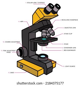
Assignment 3 microscope - Care and Structure of the Compound ...

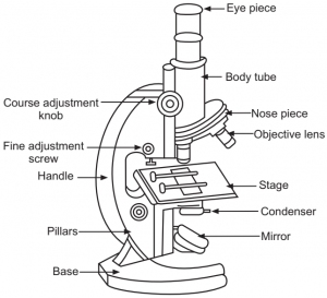
Can someone can send me diagram of this compound microscope ...

What is a Compound Microscope? | Microscope World Blog
What is a Compound Microscope? | Flinn Scientific
2,024 Compound Microscope Stock Photos, Images & Photography ...

Compound Microscope- Definition, Labeled Diagram, Principle ...

HOw to draw light or compound microscope step by step / Microscope diagram

Parts of a microscope with functions and labeled diagram

Compound Microscope Parts, Functions, and Labeled Diagram ...

Microscope With Labels Clip Art at Clker.com - vector clip ...
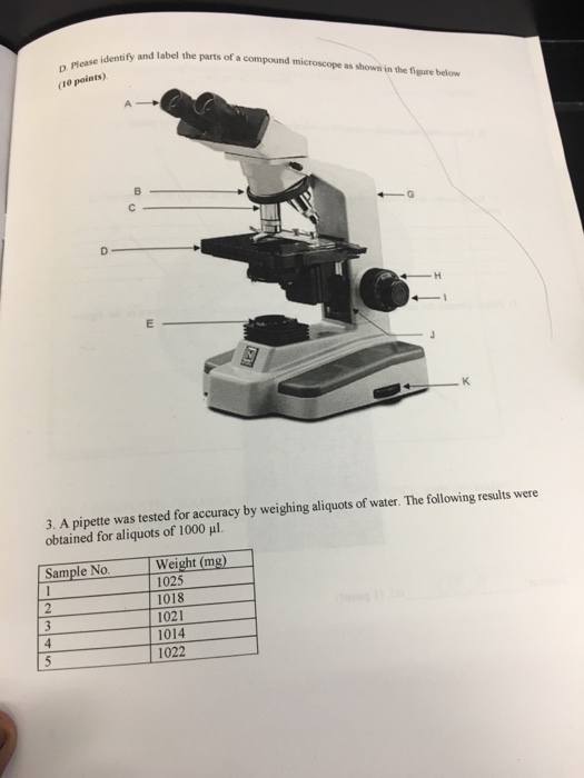
Solved Identify and label the parts of a compound microscope ...

Compound Microscope Labeled Diagram | Quizlet

microscope | Types, Parts, History, Diagram, & Facts | Britannica

Microscope Diagram Labeled, Unlabeled and Blank | Parts of a ...

Celestron Labs CM400 Compound Microscope | Celestron

OMAX 40X-2500X Trinocular Biological Compound Microscope with Replaceable LED Light

Compound Microscope Parts – Labeled Diagram and their ...
Label the Parts of a Compound Light microscope - BIOLOGY JUNCTION
Parts Of A Microscope - Parts Of A Compound Microscope, HD ...
3,469 Compound Microscope Images, Stock Photos & Vectors ...

Lasec Education | Key parts of a compound microscope and how ...

Compound Microscope Diagram | Quizlet

Compound Microscope Parts, Diagram Definition, Application ...

Microscope Diagram Labeled, Unlabeled and Blank | Parts of a ...
What is Compound Microscope? - Diagram, Function, Advantages

Parts of a Microscope - SmartSchool Systems

The Compound Microscope parts & how ... | Microscope parts ...

Compound Microscope Parts – Labeled Diagram and their ...

Compound Microscope: Know Definition,working, diagram, properties

This is a common compound microscope. Label its parts from A ...
16 Parts of a Compound Microscope: Diagrams and Video ...
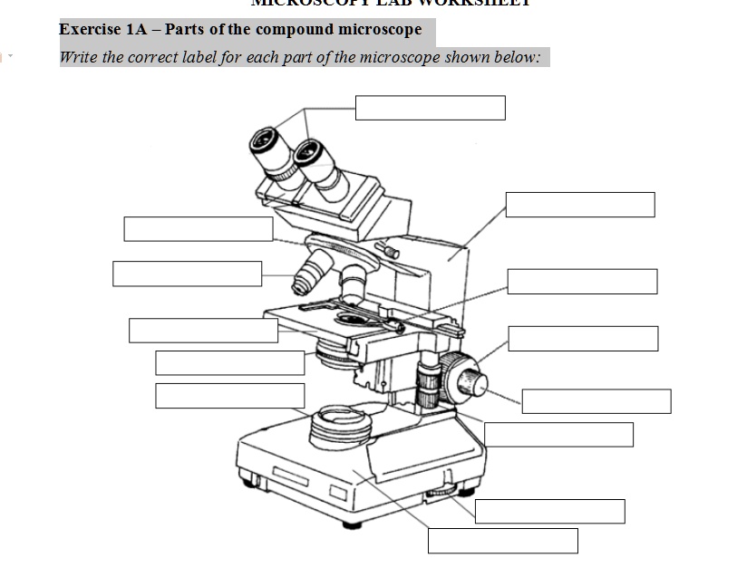
SOLVED: Exercise 1A Parts ofthe compound microscope Write the ...
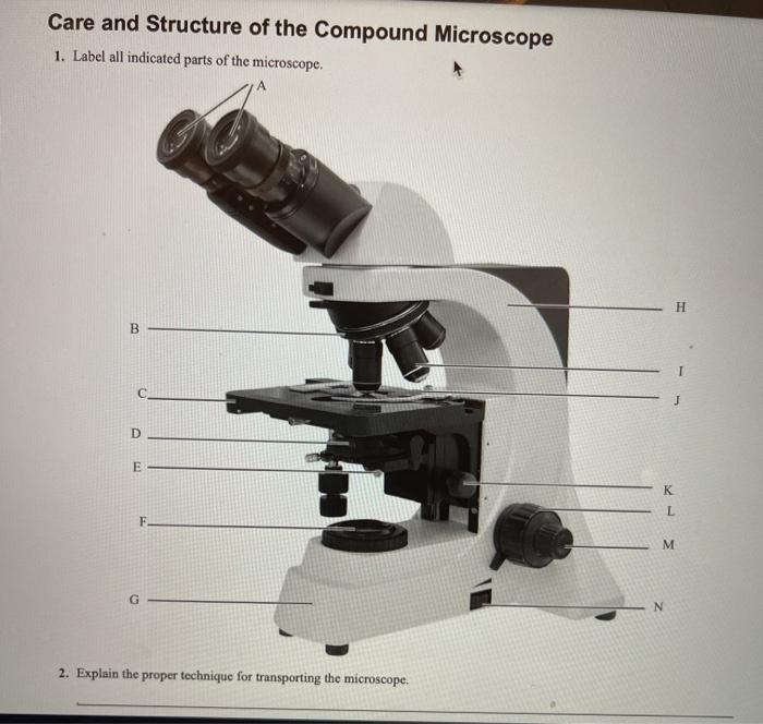
Solved Care and Structure of the Compound Microscope 1 ...
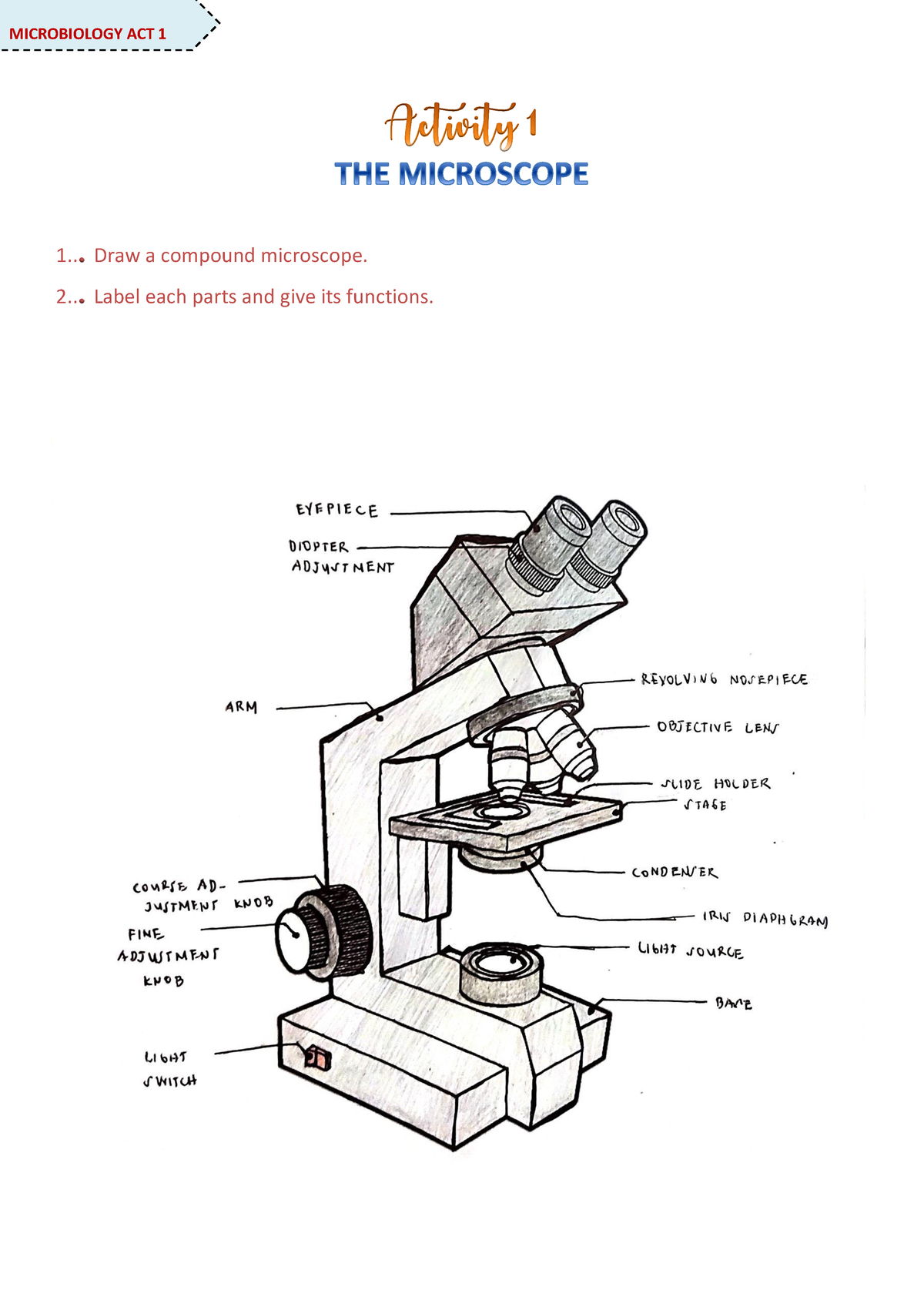
Microscope Activity - MICROBIOLOGY - 1... Draw a compound ...

Labeling the Parts of the Microscope | Microscope activity ...

Compound microscope - their parts and function - Microscopy4kids
Solved Nikon Parts of the compound microscope Write the ...

This is a common compound microscope. What the labelling D ...

Compound Microscope Parts, Functions, and Labeled Diagram ...

National 131-RLED-MS Compound Microscope with Mechanical Stage
9.1: Using Microscopes - Biology LibreTexts
Post a Comment for "41 picture of compound microscope with labels"